

Digital Diagnostics in Dentistry
31-10-2024In the field of dentistry, digital diagnostics represents a set of technologies that are widely utilized by dental professionals to provide the most accurate assessment of patients' oral and dental health. These methods offer more precise images and information, thereby enhancing the accuracy and effectiveness of treatments. The most important digital diagnostic technologies are viziography, 2D panoramic X-ray, and 3D tomography. Each technology has its own distinctive advantages, operating principles, and areas of application.
This article will provide a comprehensive overview of the three main digital diagnostic methods, examining their role in dental practice, impact on patient care, and how they simplify the work of dentists. The advent of digital diagnostics represents a pivotal turning point in modern dentistry, ushering in greater efficiency and safety.
Viziography: The Benefits of Digital Dental X-rays

Viziography is a system that replaces traditional X-ray technologies in dentistry with modern digital diagnostic methods. Compared to traditional X-ray techniques, viziography typically provides more accurate and detailed images while significantly reducing radiation levels. This offers numerous advantages for both patients and dentists. The modern approach of viziography allows for faster and more accurate results during dental examinations.
How Viziography Works
In the viziography process, digital sensors are used instead of traditional X-ray films. These sensors transmit the image instantly to a computer, where the images are immediately ready for analysis. While traditional X-ray films take longer to develop, viziography images are obtained within seconds. This speed allows dentists to make quicker decisions. Unlike traditional X-ray films, digital images can undergo various analyses and adjustments. For example, the image can be zoomed in, contrast levels can be changed, and filters can be applied to enhance specific areas, ensuring a more accurate diagnosis.
Advantages of Viziography
- Faster and more accurate diagnostics: With digital imaging technology, dentists can diagnose more quickly and accurately. Since images are obtained instantly, a treatment plan can be prepared quickly.
- Reduced radiation exposure: Viziography exposes patients to significantly less radiation than traditional X-rays, which is especially important for children, pregnant women, and patients who need frequent imaging.
- Real-time imaging: Viziography provides real-time images, which immediately enhances diagnostic capabilities. Dentists can make necessary adjustments to the image instantly, optimizing the treatment process.
- Detailed images: The images obtained with viziography are more detailed. Even small cavities, gum problems, and other pathologies can be detected in their early stages, facilitating timely and easier treatment.
Easy sharing: Digital images can be stored on a computer and easily shared with other specialists for a second opinion. These images can also be kept for a long time, making it easy to refer back to them when needed in the future.
2D Panoramic X-ray: A Comprehensive View of Oral and Jaw Structures

2D Panoramic X-ray provides a comprehensive view of the mouth and jaw. Dentists use panoramic X-rays extensively to get a full picture of the jaw, teeth, and surrounding tissues. This technology is particularly useful when multiple issues need to be analyzed at once across a wide area. The 2D panoramic X-ray allows dentists to make more informed decisions during examinations.
Diagnosis of Oral and Jaw Problems
The 2D panoramic X-ray enables a full assessment of the condition of the jawbones, teeth, and other oral structures. This imaging method reflects fractures in the jaw, cracks in the bones, and other structures, helping dentists make a more accurate diagnosis. The alignment of teeth, orthodontic problems, and other pathologies are also clearly visible with panoramic X-rays. Gum diseases, problems in tooth roots, and infections around the roots can be more easily detected with this technology.
Usage in Orthodontic and Surgical Planning
In orthodontic treatments, the 2D panoramic X-ray plays a crucial role, as it provides a broad view to evaluate the correct alignment of the teeth and jaw. Orthodontists use these images to understand the relationship between the jaw and teeth accurately. In surgical procedures, the panoramic X-ray plays an important role in planning by assessing the condition of the jawbones and the positioning of the teeth. Any anomalies in the jawbone, tooth positioning, or tooth extraction are key insights provided by the panoramic X-ray.
Analysis of Tooth Roots
The 2D panoramic X-ray is not only effective for analyzing teeth but also for evaluating the condition of the tooth roots. The health of tooth roots is as critical as the tooth itself because the roots anchor the tooth to the jaw. This diagnostic method assists dentists in the following cases:
- Root infections and inflammations: It helps detect inflammation or infection around the tooth roots in their early stages.
- Root resorption: It provides diagnostic support in cases where the tooth roots dissolve or are damaged over time.
- Root canal treatment planning: It offers precise information about the condition of the tooth roots during endodontic treatment, i.e., root canal therapy.
- Monitoring root development: This method is ideal for tracking the development stages of tooth roots, especially in children and adolescents.
Thanks to this digital diagnostic method, dentists can detect root problems at an early stage, applying appropriate treatment plans. The 2D panoramic X-ray plays an essential role in various branches of dentistry and significantly contributes to patient health.
3D Tomography: Detailed and Three-Dimensional Imaging

3D tomography is one of the most advanced diagnostic technologies in dentistry. It provides three-dimensional images of the teeth and jawbones, making it widely used, particularly in surgical and orthodontic interventions. 3D tomography helps dentists better understand jaw structures, simplify the diagnostic process, and optimize treatment plans.
How 3D Tomography Works
3D tomography technology creates comprehensive images of the jaw and teeth using X-ray beams. These images allow dentists to clearly see the exact positioning of the jaw and teeth, nerve pathways, and other vital structures. The system uses computer programs to analyze the collected data and produce three-dimensional images. This process allows dentists to make more reliable decisions from the start.
Advantages of 3D Tomography
The greatest advantage of 3D tomography is its sensitivity to detail. Compared to traditional 2D images, this technology provides a complete view of the teeth and jawbones. For example, before placing dental implants, 3D tomography can be used to determine the exact location for implant placement. 3D tomography also emits less radiation, which is crucial for patient health by minimizing radiation risks in such imaging procedures.
Detailed Analysis for Dental Implants

3D tomography provides a complete three-dimensional view of the jaw structure before dental implants are placed. This allows dentists to plan the placement of implants more accurately. It provides detailed information about the height, thickness, and density of the jawbones. This information is crucial in determining the best locations for dental implants. Additionally, it shows the precise location of nerves and blood vessels, enabling dentists to make decisions that avoid damage to these structures.
Use in Orthodontic Planning and Treatment

3D tomography is a critical imaging method in the orthodontic treatment process. The three-dimensional images of the teeth and jaw provide precise data for treatment planning. Orthodontists use 3D images to ensure correct teeth positioning, prepare treatment plans, diagnose jaw problems, and monitor treatment progress. This allows for personalized approaches tailored to each patient’s needs.
Detection of Trauma and Jaw Problems
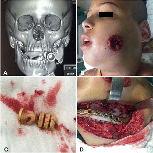
3D tomography is a modern tool for diagnosing trauma and jaw issues. These images are used to detect fractures and cracks in the jaw during jaw trauma. Additionally, the condition of the teeth and surrounding tissues is important in cases of dental trauma. The 3D images are widely used to identify the structure and condition of the jawbones, such as diagnosing problems related to temporomandibular dysfunction (TMD). These images are also obtained post-treatment to track recovery, helping adjust the treatment plan.
This structure ensures that each subsection maintains the same logical framework and volume. Thus, the importance of 3D tomography in dentistry is presented more clearly and systematically.
Conclusion
Digital diagnostics in dentistry is a modern and effective approach to accurately assessing patients’ oral and dental health. Viziography, 2D panoramic X-ray, and 3D tomography provide dentists with more detailed and accurate images, simplifying the diagnosis and treatment process. These technologies reduce patients' radiation exposure, provide real-time images, and accelerate treatment.
By using these modern technologies, dentists can detect patients’ problems earlier, prepare more precise treatment plans, and develop personalized approaches tailored to each patient's needs. Ultimately, digital diagnostics enhances patient satisfaction in dentistry and ensures more successful treatment outcomes. In the future, the advancement of these technologies will continue to drive progress in dental practices and help protect patients’ health.